

Jiao’s Scalp Acupuncture
Jiao Shunfa, drawing on clinical experience and the principle of functional localization in the cerebral cortex, developed the system of Jiao’s Scalp Acupuncture. By mapping cortical functional areas—such as the motor, sensory, and language regions—onto the scalp surface, he defined specific therapeutic zones for needle stimulation. This approach has demonstrated significant clinical effectiveness in treating neurological conditions such as stroke, paralysis, and aphasia.


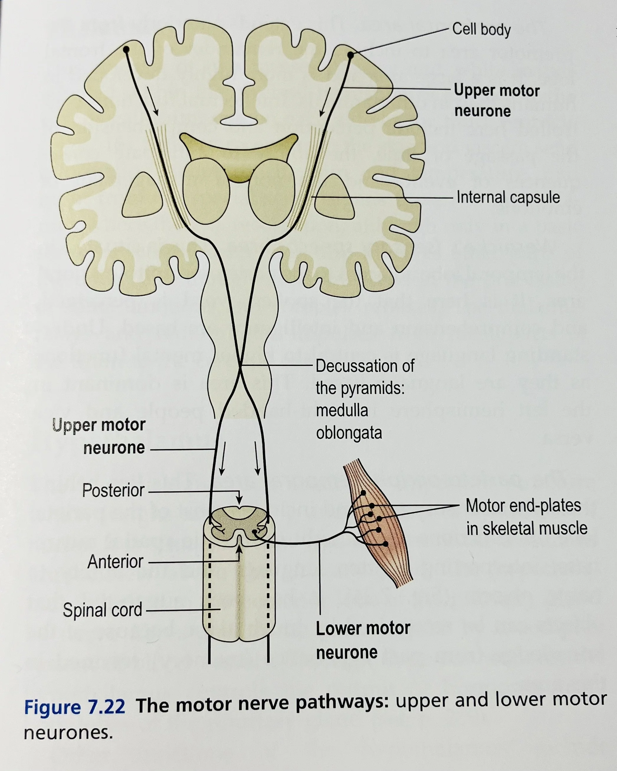
Brain Anatomy & Physiology
Chinese Scalp Acupuncture content is based on the Anatomy & Physiology of the brain


Jiao’s Scalp Acupuncture
Jiao Shunfa, drawing on clinical experience and the principle of functional localization in the cerebral cortex, developed the system of Jiao’s Scalp Acupuncture. By mapping cortical functional areas—such as the motor, sensory, and language regions—onto the scalp surface, he defined specific therapeutic zones for needle stimulation. This approach has demonstrated significant clinical effectiveness in treating neurological conditions such as stroke, paralysis, and aphasia.


T Point (Top of Motor Line / Scalp Point T)
The T point is located at the midpoint between the apices (tips) of both ears,
or alternatively, it can be defined as the midpoint between the glabella (the area between the eyebrows) and the external occipital protuberance (inion).
—————
Motor Area (Scalp Acupuncture) – Location and Indications
Upper point of the Motor Area: Located 0.5 cm posterior to the midpoint of the midline of the head (point T).
Lower point of the Motor Area: Located at the anterior border of the temporal hairline (temporal notch).
Indications by Motor Area Sections:
Upper 1/5 of the Motor Area:
➤Used to treat paralysis of the lower limb and trunk.Middle 2/5 of the Motor Area:
➤ Used to treat paralysis of the upper limb and trunk.Lower 2/5 of the Motor Area (also called Speech Area I):
➤ Used to treat facial paralysis, aphasia, drooling, and speech disorders.
—————
Sensory Area (Scalp Acupuncture) – Location and Indications
Location of the Sensory Area:
Upper point: Located 2 cm posterior to the T point (which is also 1.5 cm posterior to the upper point of the Motor Area).
Lower point: Located at the anterior border of the temporal hairline (temporal notch).
This line runs parallel and posterior to the Motor Area.
Indications by Section:
Upper 1/5 of the Sensory Area
➤ Treats:
Sensory disturbances of the lower limbs, trunk, and head
Numbness, paresthesia
Head and neck pain, dizziness, tinnitus
Middle 2/5 of the Sensory Area
➤ Treats:
Upper limb sensory dysfunction, such as:
Pain, numbness, paresthesia
Lower 2/5 of the Sensory Area (also called Speech Area I)
➤ Treats:
Facial numbness
Migraine
Temporomandibular joint disorders (TMJ)
_______
Dizziness and Auditory Area – Location and Indications
📍 Location:
Starts 1.5 cun above the ear apex (Erjian)
Extends horizontally 2 cm forward and 2 cm backward, forming a 4 cm horizontal line centered above the ear apex
📌 Indications:
Dizziness (vertigo)
Tinnitus
Hearing loss
___________
Vasomotor Area - Location and Indications
📍 Location:
Located 2.5 cm anterior to the T point
(or 3 cm anterior to the upper point of the Motor Area).
It runs parallel and anterior to the Motor Area, more forward than the Chorea and Tremor Control Area.
📌 Indications:
Primary hypertension
Cortical edema (cerebral or peripheral edema)
—————-
Chorea and Tremor Control Area – Location and Indications
Location:
This area starts 1 cm anterior to the T point
(or 1.5 cm anterior to the upper point of the Motor Area).
It runs parallel and anterior to the Motor Area.
Indications:
Chorea (e.g., Huntington’s disease)
Parkinsonian tremor
Parkinsonism syndromes
————-
Speech Area II – Location and Indications
📍 Location:
Located 2 cm inferior (downward) to the parietal tubercle
From that point, draw a 3 cm vertical line downward
📌 Indications:
Nominal aphasia (anomic aphasia) — difficulty in naming objects
Patients understand the function of an object but cannot recall its name
🧪 Clinical Example:
A patient sees a chair and knows it's for sitting, but cannot say "chair" — only says "sit"
_______
Speech Area III – Location and Indications
📍 Location:
Start from the midpoint of the Dizziness and Auditory Area (i.e., 1.5 cun above the ear apex)
Draw a 4 cm straight line posteriorly (toward the back of the head)
📌 Indications:
Sensory aphasia (Wernicke’s aphasia)
Patient has difficulty understanding spoken language
May respond inappropriately or speak fluently but meaninglessly
🧪 Clinical Example:
When asked “What is your name?”, the patient may reply with an unrelated answer
Appears fluent but cannot comprehend the question properly
——————
Practice Area (Apraxia Area) – Location and Indications
📍 Location:
Draw a line from the parietal tubercle (parietal eminence) to the mastoid process
Then draw two lines at a 40° angle in front of and behind this reference line
→ These angled lines represent the Practice Area boundaries
📌 Indications:
Apraxia (inability to carry out purposeful movements despite normal strength and coordination)
Disorders involving:
Muscle strength
Muscle tone
Basic motor execution
Motor planning and fine motor skills
___________
Lower Limb Motor-Sensory Area – Location and Indications
📍 Location:
Connect the upper points of the Motor Area and Sensory Area with a straight line
From this line, move 1 cm laterally (to the left and right sides)
From each lateral point, draw a line 3 cm in total length (1.5 cm forward, 1.5 cm backward)
The line runs parallel to the midline of the head
📌 Indications:
Paralysis, numbness, or pain in the contralateral lower limb
Acute lumbar sprain
Nocturia, urinary frequency
Uterine prolapse
_______
Visual Area – Location and Indications
📍 Location:
On the horizontal line at the level of the external occipital protuberance (枕外粗隆)
1 cm lateral to the midline (on both left and right sides)
From that point, draw a 4 cm straight line upward, parallel to the midline of the head
📌 Indications:
Cortical visual impairment
e.g. visual field loss or decreased vision due to occipital lobe dysfunction
Common in stroke, trauma, or cerebral ischemia
———————
Balance Area – Location and Indications
📍 Location:
Starting at the external occipital protuberance (粗隆水平線) level
3.5 cm lateral to the midline on both sides
From that point, draw a 4 cm straight line downward, parallel to the midline
📌 Indications:
Cerebellar diseases causing:
Ataxia (coordination disorder)
Balance problems
Dizziness (vertigo)
Brainstem dysfunction causing:
Limb numbness
Paralysis
___________
Stomach Area – Location and Indications
📍 Location:
Starting at the hairline directly above the pupil
Draw a 2 cm straight line upward, parallel to the anterior-posterior midline of the head
📌 Indications:
Gastritis
Gastric ulcer
Symptoms such as epigastric pain and upper abdominal discomfort
______
Thoracic Cavity Area – Location and Indications
📍 Location:
Medial to the Stomach Area, between it and the midline
At the level directly above the inner canthus (inner corner of the eye)
From the hairline, draw a 2 cm line upward and a 2 cm line downward, forming a 4 cm vertical line in total
The line is parallel to the anterior-posterior midline
📌 Indications:
Bronchial asthma
Chest discomfort
Respiratory-related disorders
————-
Genital Area – Location and Indications
📍 Location:
Start directly above the outer canthus (outer corner of the eye)
From the frontotemporal angle of the hairline (額角處), draw a 2 cm vertical line upward, parallel to the anterior-posterior midline
📌 Indications:
Disorders of the reproductive organs
Uterine bleeding
Pelvic inflammatory disease (PID)
Uterine prolapse
焦順發 頭針
焦順發根據臨床及大腦功能定位原理
Top T : 兩耳尖中點 或 (眉間 和 枕外粗隆 之中間點為T點)
運動區
定位
運動區上點 在 T 向後 0.5cm
運動區下點 接鬂角前缘
主治
運動區上部 : 上1/5 為下肢軀幹癱瘓
運動區中部 : 中2/5 為上肢軀幹癱瘓
運動區下部(又稱言語1區) : 下2/5 為面癱, 失語, 流涎, 發音障礙
_______
感覺區
定位
感覺區上點 在 T 向後 2cm (在運動區向後 1.5cm)
感覺區下點 接鬂角前缘
主治
感覺區上部 : 上1/5 為下肢感覺, 軀幹, 頭, 麻木, 感覺異常, 頭頸痛, 頭暈, 耳鳴
感覺區中部 : 中2/5 為上肢感覺, 痛, 麻木, 感覺異常
感覺區下部(又稱言語1區) : 下2/5 為面麻木, 偏頭痛, 顳頷關節炎
_______
舞蹈震顫控制區
定位
在 T 向前 1cm (在運動區向前 1.5cm)
主治
舞蹈病, 震顫麻痹, 震顫麻痹綜合症
_______
血管舒縮區(治水腫)
定位
在 T 向前 2.5cm (在運動區向前 3cm)
主治
原發性高血壓及皮層性水腫
_______
暈聽區
耳尖上1.5吋前後水平伸延2cm
主治 眩暈, 耳鳴, 聽力下降.
_______
言語2區
頂骨折結節後下方2cm,引一3cm 向下直線
主治 命名性失語. 不會叫椅,只能說坐.
_______
言語3區
在暈聽區中點向後引4cm
主治 感覺性失語. (問非所答,理解語言能力障礙)
_______
運用區
頂骨結節向乳突引一直綫,與此線夾角40度前後兩線.
主治 失用症, 肌力肌張力和基本運動失常,不能扣鈕,拾硬幣.技巧障礙.
_______
足運感區
運動區和感覺區上點連一直線, 左右旁開1cm, 向前後伸延共3cm, 平行正中線
主治 對側下肢癱瘓, 麻木, 疼痛, 急性腰扭傷, 夜尿, 尿頻, 子宮下垂.
_______
視區
粗隆水平線上, 中線旁開1cm, 向上引4cm長直平行線
主治 皮層性視力障礙
_______
平行區
中線旁開3.5cm, 粗隆水平線向下引平行線長4cm
主治 小腦疾病引起的共濟失調、平衡障礙、頭暈、腦幹功能障礙引起的肢體麻木癱瘓
_______
胃區
瞳孔直上頭髮制為起點,向上引線,平行於前後中線2cm長
主治 胃炎、胃潰瘍 引起的位痛、上腹部不適
_______
胸腔區
(眼內角對上)
胃區與前後正中線之間, 從發際上下各引2cm長直線, 即共4cm
主治 支氣管哮喘、胸部不適等
_______
生殖區
(眼外角對上)
從額角處向上引線平行於前後正中線的2cm長直線
主治 生殖器官疾病、子宮出血、盆腔炎、子宮垂脫等


Scalp acupuncture angle of insertion is 15 degree or below. 頭針多數用平刺透針方法,入針角度小於15度。
Scalp Acupuncture with Microcurrent Therapy
accelerating therapeutic efficacy
微電流療法是一種安全、無痛且有效的止痛療法
MCT5.3 Microcurrent therapy is a safe, painless, and effective treatment for pain relief.

DEVICE MCT5.3 OPERATION
LED no (L)
1 ... 0.3 Hz …… 5min … Relief 舒緩
2 … 30 Hz …… 10min … Pain killer 止痛
3 … 300 Hz (淺層)
4 … 7.8 Hz
5 … 930 Hz …shallow (淺層)
LED no (R)
1 … 1.7 Hz …… no change 不變
2 … 3.9 Hz
3 … 300 Hz … shallow (淺層)
Operation 操作 for 7 different purpose:
1
Ionization Bath 浸浴黑膠
Immune system & skin disease
Bath pad 黑膠
L4 + R2 浸浴 bath 20-30 min
2
silicon pad 傷痛貼 / rod 傷痛棒
Pain (when using silicon pad 矽膠貼 or rod棒)
Step 1 …. L3 + R1 for 2 min (option)
Step 2 …. L2 + R1 for 2 to 10 min
Step 3 …. L1 + R1 for 2 to 5 min
3
Acupuncture 針灸
Step 1 …. L3 + R3 for 5 min
Step 2 …. L5 + R1 for 4 min
Step 3 …. L3 + R1 for 3 min (option)
Step 4 …. L2 + R1 for 2 min
Step 5 …. L1 + R1 for 1 min
4
Lymphatic Boost 循環系統
(when using silicon pad矽膠貼)
Step 1 …. L2 + R1 for 15 min
(L)KI1 (R)PC8 左湧泉 右勞宮 (即左腳底 右手掌心)
5
EYE 眼
(when using rod棒 with wet Q-tips)
Step 1 …. L2 + R1 for 1 min
Step 2 …. L1 + R1 for 1 min
穴位組 point:BL1 & GB1 睛明穴 瞳子髎穴 / EX-HN4 & ST1 魚腰穴 承泣穴 / SJ23 & BL2 絲竹穴 攢竹穴
6
NOSE 鼻
(when using rod棒 with wet Q-tips )
Step 1 …. L2 + R1 for 1 min
Step 2 …. L1 + R1 for 1 min
穴位組 : (L&R)LI20 左右迎香穴
7
FACE 面部
Face treatment
(when using rod棒 with wet Q-tips)
Step 1 …. L3 + R1 for 1 min (option)
Step 2 …. L2 + R1 for 2 min
Step 3 …. L1 + R1 for 1 min


方雲鵬頭針
以伏象、倒象、伏臟、倒臟為主。


方雲鵬頭針
以伏象、倒象、伏臟、倒臟為主。


